By Steve Fisch, DVM
The main goal of the respiratory system is to transfer oxygen from the air that is breathed to the red blood cells where the oxygen will be transported throughout the body. In addition, carbon dioxide, a waste product of metabolism, is eliminated from the body via the lungs. If this system is compromised in any way it will severely affect the athletic ability of a horse and may even threaten his life. Ultrasound is one diagnostic tool that can aid in the diagnosis of pleuritis and pneumonia.
The lower airway consists of the lungs and bronchi. The lungs have protective mechanisms that help prevent infection. The air we breathe is not sterile and contains many contaminants such as dirt, dust, pollen and chemicals, as well as bacteria, viruses and fungi.
Protection from such contaminants actually starts in the upper airway with filtering, humidifying, and warming of the inspired air. The upper respiratory system, trachea, and bronchi are lined with tissue that is always covered with sticky mucus that traps contaminants. The tissue lining the trachea and bronchi has cells with extremely small hair-like fibers sticking out into the airway called cilia. The cilia increase the surface area of the filtering surface and play an active role in airway protection. The debris that is collected on the surface of the airways is transported away from the lungs by these cilia. The small cilia fibers move in a coordinated rhythm to move the thin layer of mucus that floats on top of the cilia and the debris that is stuck to it out of the lungs and up the trachea where the horse will swallow it.
Threats to equine respiratory health
Anything that decreases the effectiveness of this process increases the chance for the development of respiratory disease. Factors such as cold air, smoke or chemicals in the air and inflammatory conditions of the airways such as COPD and flu can decrease the cilia defense mechanism. Chemicals like ammonia fumes from poorly cleaned stalls or bad barn ventilation are a common cause of decreased ciliary action.
Another important way by which the mucociliary clearance mechanism can be overwhelmed and rendered less effective is not allowing the horse to lower its head during transport. In fact, the transportation process can stress the defense mechanism of the lungs in several ways. The exhaust fumes from a poorly maintained or designed truck can have a negative impact on the cilia’s effectiveness. In addition, a trailer that has poor drainage can allow for the buildup of ammonia fumes. If the ventilation within the trailer is not good, it can make all of these negative factors threatening lung health worse.
There have been several scientific papers evaluating the effects of transportation and head posture on lung health. It has been demonstrated that horses confined with their heads elevated for 24 hours developed an accumulation of inflammatory airway secretions that was associated with increased numbers of bacteria in the lower respiratory tract. These findings indicate that hauling horses in a manner which prevents them from lowering their heads during transportation and cross-tying may contribute to lower respiratory tract disease.
There are other mechanisms in place to protect the respiratory system against infection. There are millions of special cells deep within the tissues of the lung that can kill bacteria and inactivate viruses. These cells are neutrophils and macrophages that basically eat the bacteria and viruses.
Diagnosis and Treatment 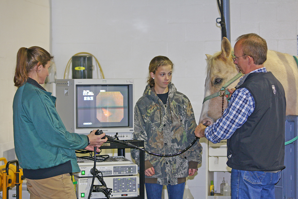 Dr. Fisch takes chest radiographs and an endoscopy of the upper respiratory tract.
Besides blood work, ultrasound, radiography, endoscopy and respiratory fluid analysis, there are ways to evaluate the respiratory system, which include evaluating respiratory rate and the horse’s character of breathing, A physical examination is required to adequately evaluate the respiratory system. It should be determined if there a nasal discharge, and if so, is it a clear and thin or thick and green/yellow discharge? Is it bloody, foul smelling, and does it come from one or both nostrils? Is the airflow from both nostrils normal? Has the horse been coughing, and if so, is the cough wet or dry? Are there any painful swellings around the head? Does the horse make a noise while breathing at rest, at work, on inspiration or expiration? Is there an extra abdominal movement while breathing? Is there a fever? Are there any other horses on the farm with similar signs?
All of these questions are used by your veterinarian to help determine the type and severity of affliction that the horse may have.
Viral Pneumonia  This foal receives hyper-immune plasma for treatment of pneumonia.
Many of the viral entities affecting the lungs also affect the upper airway. Some of these are Equine Influenza, Equine Herpes Virus and Equine Viral Arteritis (EVA).
Equine Influenza. The equine flu viruses contain a chemical that damages the mucociliary transport mechanism and allows for evasion of the immune system. The damage to the mucociliary transport system can take several days or longer to repair. This leaves the defense mechanisms decreased and exposes the horse to the possibility of secondary bacterial infection. The disease can be extremely contagious, especially in conditions of crowding and poor ventilation.
Influenza most commonly affects 2- and 3-year-olds. Stresses on the respiratory tract’s immune system and poor vaccination programs are predisposing factors. The incubation period is one to three days. The virus usually affects the upper respiratory tract more than the lungs. Clinical signs usually appear three to five days following exposure to the virus. The clinical signs include fever, anorexia, depression, a clear nasal discharge, and a deep, dry cough. The course of infection is typically from two to 10 days if there are no complications. Secondary bacterial infection is a common complication of equine influenza. Horses can shed virus for three to six days after the last signs of illness and should be kept in isolation for that period of time.
The treatment generally involves symptomatic care and the use of antibiotics only if a secondary bacterial infection is suspected. One of the greatest risks is putting an affected horse back in work too soon. The virus causes a significant amount of tissue damage, which requires time to regenerate and heal. If the horse is placed back to work too soon, it is highly likely that complications will develop. In some horses with severe infections, there might be the need for one to two months of rest prior to resumption of training. A recovering horse should be rested at least 21 days past the last sign of infection. Young foals can suffer more severely from equine influenza. They can develop extensive and fatal pneumonia.
Regular vaccination can significantly reduce the population at risk and is something I recommend. For younger athletic horses, which might be at greater risk, it is suggested that they be vaccinated at three- to four-month intervals as opposed to older horses, which should receive their boosters at six to 12 month intervals.
Equine Herpesvirus (Rhino). Equine herpesvirus, equine rhino, rhino, and rhinopneumonitis are all names for the equine herpesvirus. Rhino means the upper airway and rhinopneumonitis means inflammation of the upper airways and lungs. There are four currently known strains of the equine herpesvirus known as 1, 2, 3, and 4. It is strains 1 and 4 that are associated with respiratory disease in the horse.
Respiratory disease related to the herpesvirus most commonly occurs in foals, weanlings, and yearlings. The immunity to herpesvirus infection is short-lived and re-infection is thought to reoccur. In broodmares, there can be abortion related to infection. The equine herpesvirus type 1 has the ability to cause respiratory disease, abortion, and neurologic disease. Foals can be born suffering from extensive pneumonia and die within 72 hours.
The infection occurs via the inhalation of the virus. The virus does not survive well outside of the body for extended periods of time. Close contact with an infected animal or tissues is an important part of transmission. The clinical signs appear one to three days following infection and include fever, anorexia, depression, a clear nasal discharge, and a deep dry cough that cannot be distinguished from influenza.
The treatment is largely supportive and a convalescent period of rest is extremely important for reducing the complication rate. Vaccination will not always prevent the disease, but can reduce the severity of the disease. Pregnant mares should be vaccinated to prevent abortion.
Equine Viral Arteritis. The equine arteritis virus causes inflammation of the blood vessels. Transmission of this virus can occur via inhalation or sexual contact. The virus rapidly spreads throughout many of the body’s organs and the disease can produce signs of respiratory distress. The disease also can cause abortion.
The incubation period is three to 14 days. The main clinical signs include fever, loss of appetite, depression, and possibly a cough. A clear nasal discharge, a bright reddening of the nasal and ocular tissue, and excessive tear production can also be associated with EVA.
The treatment consists mainly of supportive therapy and treatment for any secondary bacterial infection. Rest is very important. Isolation should be maintained for three to four weeks past the last observation of clinical signs to prevent transmission. Most horses recover uneventfully but there can be fatalities.
Viral Diagnosis 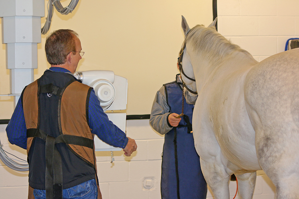 Dr. Fisch says that evaluation of respiratory distress in its early stages is advisable.
The clinical signs for most of the viral respiratory diseases are very similar. In many cases, the exact cause does not have a significant impact on the treatment, but knowing the specific cause can provide information on how to prevent the disease. A nasal swab can be acquired for an attempt to identify the virus via DNA in the laboratory. This is the most common way to make a diagnosis. In addition, there are several laboratory techniques used in identifying a virus present in a lab sample. Another diagnostic tool is to evaluate the concentration of antibodies in the blood for the suspected virus.
Bacterial pneumonia
There is a constant source of bacteria that could potentially invade the lung. There are many types of bacteria that have been identified as being involved in pneumonia in the horse. Most of these bacteria are either in the environment of the horse or are a normal inhabitant of the upper airway or throat area. Bacterial pneumonia often follows viral pneumonia due to the damage to the normal protective mechanisms and disruption of the local immune system. This situation is often the result of not allowing for an adequate rest period following a viral respiratory disease. Other stressful events that can lead to the development of bacterial pneumonia include any intense athletic exercise, transportation, poor nutrition and overcrowding.
The clinical signs associated with bacterial pneumonia include fever, depression, poor appetite, nasal discharge, coughing, respiratory distress and the presence of abnormal lung sounds. In more chronic cases, the onset can be slow and vague, with exercise intolerance and weight loss being the main clinical signs.
In some cases, the infection might be localized and walled off in the form of an abscess such as with Rhodococcus Equi in foals. If the development of the abscess is slow enough, the first recognition of the disease can be severe acute respiratory distress during exercise or a foal that is acutely depressed and febrile. The clinical signs of the disease occur when the lung abscess ruptures or leaks possibly from the stress of exercise.
The diagnosis of bacterial pneumonia often is made based on history, clinical signs, and lung sounds. Ultrasound and radiographs might be necessary to assess completely the extent of the infection. A transtracheal wash can provide important information and may help direct therapy. The treatment of bacterial pneumonia generally consists of long-term antibiotic therapy, any supportive therapy necessary, and rest. The outcome of bacterial pneumonia can be extremely variable and depends on the causes, duration of infection, amount of lung tissue involved, the specific types of bacteria involved, and the existence of additional complications.
Shipping Fever
With respect to the respiratory system, there are a number of known factors that can predispose a horse to respiratory disease.
There have been several studies evaluating the direct effects of transportation on the internal environment of the lungs. In one study a sample of fluid was obtained from within the trachea both prior to and immediately following transport. When compared to the pre-transport controls, the post-transport tracheal fluid contained signs of inflammation and an increased number of bacteria. The majority of these samples showed that a Streptococcus bacteria species was the most predominant bacteria found. Bacterial contamination of the lower respiratory tract occurs as a routine consequence of transportation of horses and is likely to be an important determinant in transport-associated respiratory disease. The disease “Strangles” is also commonly called shipping fever. Strangles can be prevented or at least have the affects lessened by vaccination.
Pleuropneumonia  Dr. Fisch advises a properly designed vaccination program as an important part of preventing respiratory disease.
The pleura is the thin covering of the lung or the thin lining of the thoracic cavity. Pleuritis is an inflammation of the space between the body wall and the lung. Pleuropneumonia is inflammation both within the lung and within the pleural cavity. In most cases, pleuritis is secondary or occurs in conjunction with pneumonia.
All of the factors that predispose a horse to the development of pneumonia are also thought to put it risk for pleuritis. The development of pleuritis in conjunction with pneumonia can greatly complicate and prolong treatment. A variety of bacteria can infect the pleural space. The type of individual bacteria that cause disease can have a great impact on the overall outcome.
The clinical signs of pleuritis/pleuropneumonia include fever, depression, nasal discharge, and being off feed. Pleuritis is an extremely painful disease process. These horses often have pain between the ribs. They are reluctant to walk because of the chest pain. One of the main clinical signs of pleuritis is the buildup of fluid within the thoracic cavity. This fluid muffles the lung sounds in such a way that there are no audible lungs below the fluid line. There are sounds heard with the stethoscope called pleural friction rubs. These abnormalities can be confirmed with the aid of ultrasonography. The fluid within the pleural cavity can generally be cultured and evaluated microscopically for the presence of bacteria.
The treatment of pleuritis can be very difficult and relies on long-term antibiotics, supportive therapy, and drainage of the fluid from the thoracic cavity should it become necessary. There are numerous complications that can occur while these horses are attempting to recover. If there are no complications, treatment can be successful.
COPD
The term COPD stands for chronic obstructive pulmonary disease, otherwise known as “heaves.” Other names for this disease include chronic obstructive lung disease, and lower airway inflammatory disease. This disease makes it difficult to expel the air from the lungs. Hence the “heave line” where the muscles hypertrophy from the extra effort to push the air out.
COPD is caused by a variety of allergic reactions. Hypersensitivity to dust and mold is one common cause. There appear to be two categories of affected horses. One group appears to be reacting to allergens within the barn and gets better when kept outside. Another group appears to be reacting to allergens in the pasture and gets better when kept inside. In the south many of these horses are allergic to Bahia grass.
The clinical signs include a chronic cough, a cloudy nasal discharge, and difficulty in expiring air. These horses usually take in air relatively well. Because COPD is primarily an allergic reaction without the presence of infection, there is generally no fever unless there is a secondary bacterial infection. Exercise intolerance, weight loss, and not eating are additional clinical signs. Sometimes these horses can get into trouble because they are so focused on breathing they will not take the time out from the effort of breathing to eat or drink.
The diagnosis of heaves is generally based on history and examination of the respiratory system. Sometimes other diagnostic tests, including a transtracheal washing, might be necessary to rule out other secondary problems. However, a bronchoalveolar lavage is preferred to a transtracheal wash for diagnosis of COPD. Allergy testing is another good aid in diagnosing the allergens.
The treatment of heaves involves altering the horse’s environment. If a horse is worse outside, then keep him inside, and vice versa. Often wetting of the hay or complete removal of hay from the diet is necessary, along with controlling the dust in the stall. Management changes are just as important as medical therapy. Medical therapy typically involves making sure there is no secondary bacterial infection. The main focus of therapy is to decrease the inflammation associated with the allergic reaction going on within the lungs. The drugs of choice for this are corticosteroids and/or bronchodilators. Steroids decrease inflammation and bronchodilators relieve respiratory distress by opening obstructed airways. During a severe crisis, the main problem is the constricted small airways within the lungs. Corticosteroids help the condition but suppress the immune system and can predispose to infection. Their use has also been associated with the development of founder or laminitis.
The drug clenbuterol or ventipulmin has been used for several years for treatment of COPD. It is a bronchodilator and it works well to allow the trapped air to leave the swollen bronchi. In severe cases clenbuterol and corticosteroids are used together.
EIPH
Exercised-induced pulmonary hemorrhage is a disease of athletic horses in which there is hemorrhage originating from within the lungs caused by the small capillaries breaking due to the high blood pressure resulting from exercise. The hemorrhage can be subtle enough to be seen only by microscope evaluation of a bronchial aspirate or volumous enough to be observed pouring from the nostrils.
Some additional clinical signs include exercise intolerance, respiratory distress, coughing, and excessive swallowing. The diagnosis is generally made by evaluation with the endoscope after exercise.
Treatment of EIPH involves ruling out the presence of infection. The diuretic drug Salix or furosemide is used to combat EIPH by lowering the capillary blood pressure in the lungs. Rest after an EIPH episode is an important part of treatment.
Conclusion
A healthy respiratory system is complex and vital to the life and athletic ability of the horse. Prevention is the best medicine. When a respiratory ailment does occur, quick and accurate veterinary diagnosis is paramount to a good outcome. Waiting to see what happens may cause you to see your best friend, athletic partner or even a new foal. In the case of the respiratory system distress, evaluating the problem sooner is better.
About Steve Fisch, DVM  Steve Fisch, DVM
Dr. Steve Fisch lives in Tallahassee, Fla., and is a 1982 graduate of the University of Georgia College of Veterinary Medicine. Dr. Fisch is one of the veterinarians at AVS Equine Hospital, which is a full service equine hospital specializing in lameness, surgery and reproduction. Their website is www.avsequinehospital.com.
Email comments on this article to [email protected].