
What to know about the causes and treatment options for Angular Limb Deformity in foals.
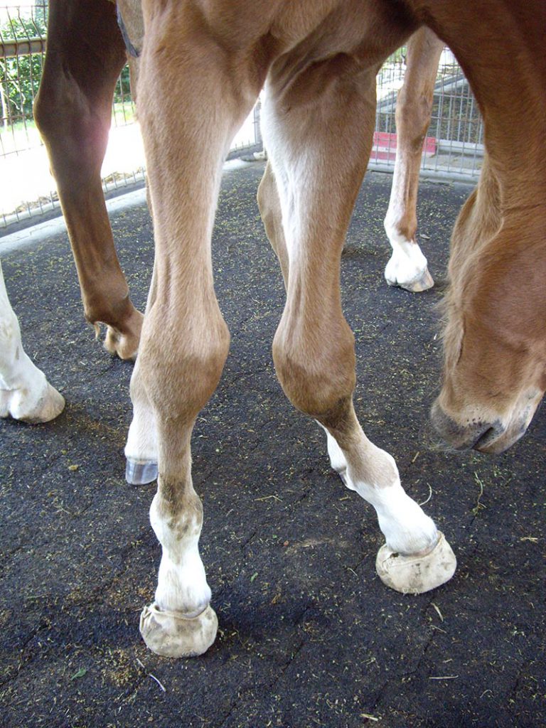
There are, however, a substantial number of cases concerning foals where Mother Nature does not lend a hand and deviation of a limb either medially (near the middle of) or laterally (on the side of) the growth plate located in the fore or hind limbs is developed or retained. This is known as angular limb deformity (ALD) and in situations where this defect may inhibit a foal’s impending soundness, a veterinarian and/or farrier may have to become involved to remedy the situation. Any animal that possesses legs longer than its head, such as a giraffe, zebra or horse, has the proclivity to leave the womb and enter this earth with legs that aren’t exactly straight. Actually, this is quite common in most foals, especially newborn Quarter Horses and Thoroughbreds, and nearly 90 percent of the time, these issues self-correct as the new arrival’s chest begins to grow outward and upward, and as he or she becomes accustomed to their new environment.
“Angular limb deformity (ALD) is a well-recognized problem in a small but significant number of foals,” wrote Dr. C.M. Colles of Avonvale Veterinary Practice, located in Oxfordshire, U.K., for the 2008 Association of American Equine Practitioners (AAEP) convention. “It is generally accepted that most cases will correct themselves with time or with careful corrective trimming and shoeing carried out before closure of the relevant growth plates. A small number of cases, however, will not straighten with conservative treatments and some corrective technique is required to prevent a conformational defect in the adult horse.”
This condition can be congenital, which means present at birth due to numerous factors including placement in the uterus prior to birth, position during the birth itself, or an inherited genetic predilection to a conformational issue. ALD can be acquired after birth, because of injury, inflammation of the growth plates, immaturity or weakness in the muscles, cartilage and ligaments of the legs, which is quite common in foals.
ALD can occur in any limb or multiple limbs and can wreck havoc with unaffected limbs as the foal may overload another limb to compensate.
“Congenital ALD recognizes different causes, including abnormal uterine positioning, hormonal or nutritional imbalances, and hereditary predisposition,” wrote Dr. F. Torre of Clinia Equina Bagnarola in Bagnarola, Italy, in a 2006 International Congress of World Equine Veterinary Association in Marrakesh, Morocco. “In these cases, ALD is present in the newborn foal. ALD deformities may also arise in association with prematurity or immaturity, when delayed ossification of cuboidal bones of the carpus and tarsus are still partially formed by cartilage, which normally completes its ossification during the last 2-3 weeks of gestation. This causes the foal to be unable to bear its proper weight without injuring its incompletely ossified bones. Bone collapse invariably follows, causing ALD.
Torre added that ALD can also be the result of uneven load distribution in cases of severe injury to the contra lateral limb, which may be affected by septic arthritis, osteomyelitis or fractures.
“In these cases, the normal limb overloaded and is prone to develop valgus or varus deformity,” he said.

So, what in layman’s terms does this actually mean? Valgus denotes a lateral or outward deviation of the affected limb below the point of the deformity, while varus is a medial or inward deviation of the same nature. The most common deviation is a knock-kneed foal, which is known medically as carpal valgus.
Differing Opinions
Surgery is not the best immediate option to relieve ALDS, no matter what the cause or site of the complication, and often limited exercise, extensions, careful trimming, glue-on shoes, casting and splinting can derive the necessary solution. If the foal has not responded to these methods by two months of age it might be the only resort.
“It becomes a question of what deformities need to be evaluated or dealt with to prevent lameness in the future. That is where you are getting a lot of differing opinions,” explained Dr. Alan Ruggles of the Rood and Riddle Veterinary Clinic in Lexington, Ky. “One side of it is there are always exceptions to the rules, meaning some horses have enjoyed tremendous athletic performance with poor conformation, and the opposite is also true.
Ruggles contends that there is more to athletic performance than conformation, however, there are certain angular deformities that are commonly associated with lameness in adult horses.
“Probably one of the most important of those is toeing in at the fetlocks, or in medical terms, fetlock varus. It’s importance is it does tend to lead to both problems in the fetlock joint itself, and secondary problems with the feet, or potentially knee or carpal deformities that we deal with in racing breeds, including barrel racers.

Ruggles cited a large group of Thoroughbred yearlings that were evaluated many years ago for conformation and followed up with for athletic performance. In this population of horses one thing associated with reduced performance was turning in at the fetlock, or fetlock varus. While fetlock varus is indeed cause for concern over a foal’s performance potential, Ruggles explains that it is still difficult to determine which foals will correct abnormalities on their own as they grown and develop, and which ones need more help.
“I would say it’s hard to get a percentage on how many foals need intervention since it depends on how intensely people manage their foals and what they are willing to accept on their own,” he continued. “But I would say that easily 25 percent of foals could use some kind of intervention. By that I mean repeated evaluations every three to four weeks. Or they may need their feet trimmed differently by the farrier, or need therapeutic extensions to help with the conformation. These 25 percent do not all need surgery and it should be performed only if there are problems with the fetlocks or carpus.”
ALD is diagnosed through routine evaluations of the foal within 24 hours of birth, several weeks after birth, and, if necessary, in the immediate months to follow. Since the canon, tibia and radial bones have differing growth rates during the first two, four and six months of life, early attention to this condition can make a tremendous impact.
“Careful clinical assessment is required to define the deformity,” wrote Dr. Mathew Smith of Newmarket Equine Hospital in Suffolk, U.K. for the 2010 British Equine Veterinary Association Congress. “This should include examination whilst standing square and with the limbs positioned straight beneath the body and when walking.
Dr. Smith emphasized that, “visual assessment should be made directly in front of the joint being assessed,” and added that radiographic examination is also critical in the assessment of ALD. For clinicians to accurately diagnose the problem, age of the foal, remaining growth potential at the site of the angular deformity, and cause and severity of the deformity must also be taken into account.
“Only after considering these factors can a suitable treatment plan be formulated,” said Smith.
Ruggles, who presented ‘Management of Angular and Flexural Disorders in Foals’ in conjunction with Dr. Wayne McIwraith of Colorado State University at the 2008 AAEP convention, stresses just how key early evaluations of the foal can be.
“In my opinion, people really need to be informed about a couple of things,” he said. “One is how good of an idea it is to have another pair of eyes look at your foal, whether it’s on one single occasion, possibly at 30 days of age, or repeatedly to correct the conformation if needed. There is a very short window of time, as you should try to have these things corrected or in the process by eight weeks to 12 weeks. There are times you have a foal where it’s growth plates close later like 12 weeks, but by then you are probably too late. Another big problem I see is people kind of forget about the feet and fetlocks, then notice them when they are yearlings. Although you can still correct the growth on the radius just above the knee to try and improve the carpal conformation, the die is already cast, because you can push the knees in some, but you can’t correct the problem. Most people don’t recognize the problem occurred six months ago and that it’s too late. Have a farrier you trust or a veterinarian evaluate the foals at 30 days, try to pick out the problem ones, take some notes and remind yourself to look at them closely again at some point.”
Surgical Options
There are three surgical options to remedy ALD. They are: periosteal stripping, also known as periosteal elevation and/or hemi-circumferential transection, transphyseal bridging, and corrective osteotomy or osteotomy.

An osteotomy is the process of removing a small piece of bone near the damaged area. The theory is that causing a shift in weight bearing from the area where cartilage is damaged to an area where it is not will encourage growth of the bone towards that spot. Everything is held in place with plates and screws. Currently, this procedure is only used if growth in the bone has ceased and there happens to be an ALD that cannot be lived with. There are two forms, the step and the wedge, with the step being the current surgical option of choice.
In periosteal stripping, which is the primary surgery utilized in ALD affecting the radius and tibia, an upturned incision resembling a T-shape is done directly above the offending growth plate at the spot where the bone is shorter on that side. The periosteum is selectively removed from the bone in order to kindle growth at that site. The Swiss veterinarian Jorg Auer introduced this surgical technique to the veterinary community around 1980 while he was at Texas A&M University.
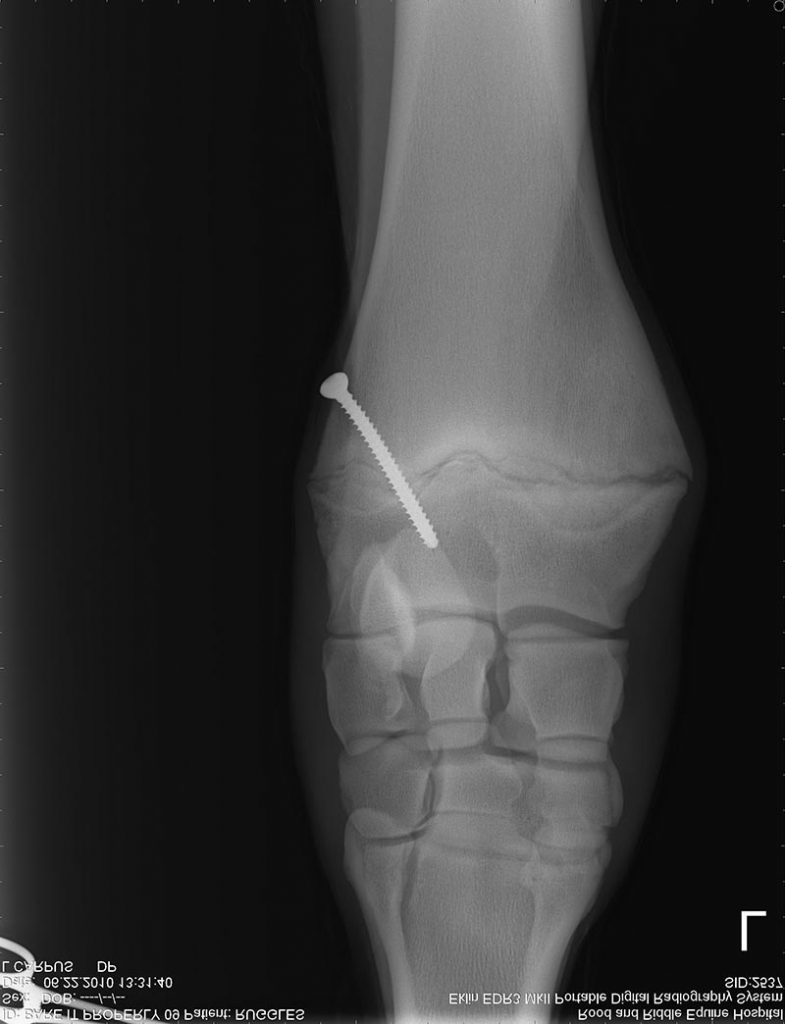
Transphyseal bridging was used before periosteal stripping arrived on the scene and is normally performed on foals less than three months old after its rapid growth rate has ceased. The goal is to slow the growth down on the medial side of the limb for a valgus deformity and on the lateral side for a varus deformity by placing pressure on the physis, or growth plate.
Until recently, there were three techniques used to create the necessary pressure upon the growth plate: staples, screws with wires in a figure 8 pattern and screws with a bone plate about 2.7mm in size. Transphyseal Screw Technique is the latest method, where either a single screw or screws with an tension band wire loop are implanted.
Although surgical repair of ALD has been around for more than 40 years and there are a multitude of papers discussing this form of correction, there is no manual so to speak for treatment. Consequently, veterinarians must rely on these considerations: the age of the foal, the site of the ALD, how bad the deformity is, and whether the deformity is congenital or acquired, improving, remaining the same or worsening.
“Really there are just two major surgeries and that’s PE (periosteum elevation) and TB (transphyseal bridging),” Ruggles said. “The short side of the bone is the part where the deformity is pointing toward, so if the foal is toed in at the fetlock, the cannon bone is the short side and the long side is the outside. We will accelerate growth by elevating the periosteum, which is the covering of the bone, to stimulate growth in the plate to help correct the deformity.
“With the other method (transphygeal bridging) growth is temporarily stopped on the long side of the bone with screws that go across the growth plate or screws in the bottom of the bone, or epiphysis, and above the bone called the metaphysis. Then they are connected with wire. These implants are then removed once the conformation improves. This is called physeal retardation and is a very effective way of correcting deformities, especially for the foals that look pretty normal until they are about eight to 10 weeks old and all of a sudden start toeing in, because as soon as you put the screw in they can’t grow anymore. At that point, periosteal elevation is not very effective, but you can absolutely stop and correct the deformity as long there is growth potential with a screw. We can do this for the carpus and tarsus (hock) as well, but there are certain ages where we do one procedure or another. There is some nuance and you really need to consult with your veterinarian to find out which technique is right at which site and how old the foal is because it really does matter.”
Ruggles emphasized that the surgeries can be successful in several circumstances, but explained that the extent of the deformity must be determined in order to know if it’s caused significant permanent deformity to the underlying bone.
“A good example of that is a foal that was knock-kneed especially badly in one leg, with badly deformed being more than 15 degrees from a straight line, because that could lead to them crushing the carpal bones, which would rupture the collateral ligaments, and then transphyseal bridging would not be successful because it doesn’t have the capability,” said Ruggles.
“People have to recognize there is a time limit,” he continued. “Deformities are not that severe if they haven’t gotten to the point where we have secondary issues. You pay attention to it early and when they have straight fetlocks at two or three months old, you can check it off the list of all the normal perinatal things you have to keep track of. There is a consequence of letting it go too long and if you don’t recognize the problems, you may lose your opportunity to correct them.”
Studies and Research on Surgical Options
There is much debate in the veterinary community over the efficacy of periosteal stripping. In 2001, Doctors Emma Read, Matt Read, Chris Clark, John Pharr and David Wilson, all from Western College of Veterinary Medicine at the University of Saskatchewan, presented ‘An Evaluation of Hemicircumferential Periosteal Transection and Elevation in an Angular Limb Deformity Model.’ For this study, 10 mixed breed foals were equipped with transphyseal bridging at 30 days of age. The implants were removed when they were 90 days old and a random limb was chosen to perform periostreal stripping with the contra limb serving as the control device. The foals also had their feet tended to throughout the study.
“The use of temporary transphyseal bridging allowed creation of a consistent angular limb deformity model that allowed us to evaluate HCPTE (Hemicircumferential Periosteal Transection and Elevation),” wrote the doctors. “In our model, the use of HCPTE as a treatment of angular limb deformities ascribed to long bone growth disparity was no more effective than stall confinement and hoof trimming alone. The transphyseal bridge placement significantly increased the angulation of both limbs with no significant difference between the limbs. By approximately eight weeks after the periosteal stripping, the angulation of both limbs had significantly improved; however, there remained no significant difference between the treated and control limbs.”
In 2000, Dr. Donnie Sloan and his team of researchers from Peterson and Smith Equine Hospital in Ocala, Fla., published ‘Restricted Exercise and Transphyseal Bridging For Correction of Angular Ligament Deformities,’ after it was presented at the AAEP convention.

Sloan essentially recreated Auer’s experiments where periosteal stripping was engaged on a normal foal in one leg on the inner side of the lower end of the radius and he determined this made the foals become knock-need on the 30th day following the procedure. When Auer did not discover any growth at the site of stripping, he assumed the resulting growth must have been in the upper end of the radius.
In the Peterson and Smith examination, Sloane concluded restricted exercise corrects nearly all congenital angular limb deformities and if this practice does not yield results, transphyseal normally remedies the condition.
“Nothing happened,” Slone, who used eight mixed-breed horses for his study, said in The Blood-Horse in January of 2002. “The foals didn’t become knock-need. No statistically significant difference between medial and lateral growth rates at either the proximal or distal end of the radius could be detected between the treated and the control limbs.”
“We don’t usually do anything drastic to their feet, but trimming is important,” he continued. “Almost any foal that is knock-kneed will turn out at their feet. They need to have the outside of their feet trimmed shorter and their toes rounded. But mostly, you just have to be patient, unless you are dealing with fetlock angular limb deformities. Periosteal stripping is something that just needs to go away. We haven’t recommended its practice for at least 10 years. We believe the procedure gets credit for correcting those angular limb deformities that would correct without intervention.”
Dr. Wayne McIwraith and Dr. Tina Anderson from Colorado State University studied a population of Thoroughbred for conformational development as weanlings, yearlings, 2-year-olds and 3-year-olds by taking three photographs from the left side, front and rear of each horse. The conformation information was documented and included with information about the horse’s future performance on track and how sound they remained.
“We found that some degree of carpal valgus protects a horse from knee injuries,” McIlwraith, who is the chair of the Orthopedic Research center at the University’s College of Veterinary Medicine, said to The Blood Horse in January of 2002. “In other words, if you have a really badly knock-kneed horse, that’s a problem. But if he’s only got a little bit of a carpal angle—less than eight degrees—that’s good. When we looked at the correlation coefficient between carpal angle and clinical problems, we found the straighter the leg, the more problems they had, so some people’s obsession at the sales with having a dead straight knee is a little bit counterproductive to racing soundness.”
Although the benefits of periosteal stripping and it’s future use in correcting ALD are in question, it seems if a horse does not autocorrect and surgery is absolutely necessary, transphyseal bridging appears to be a viable option.
Pennsylvania resident Kimberly French is a freelance writer whose work has appeared in Thoroughbred, Standardbred and Quarter Horse publications. Email comments on this article to [email protected].