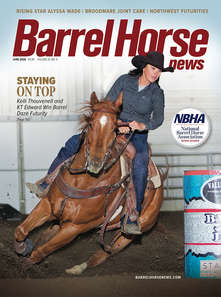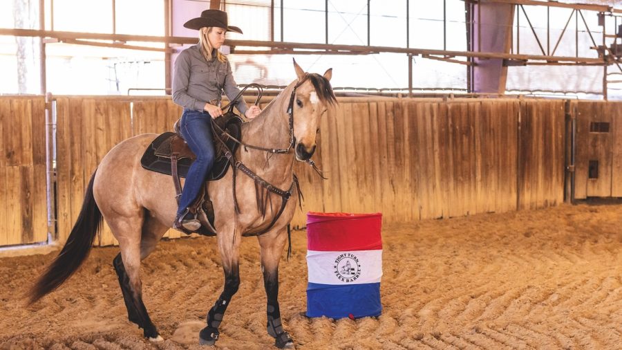
 Barrel horses can be especially prone to stifle issues due to the hard-turning nature of the sport. Photo by Abigail Boatwright. An OCD is a condition where there is a defect in the articular cartilage and the subchondral bone (bone just deep to the articular cartilage). This defect can develop in the cartilage or the cartilage and bone. The stifle (femoropatellar joint) is one of the most common places to find this lesion. If your veterinarian is suspicious of an OCD based on the information gathered from the history, physical, and moving examination, they will take radiographs of the stifle. So, once your horse is diagnosed with OCD lesions, what do you do? Based on history and presentation, there are a few routes you can take. If this is a young horse and presented with swelling of the stifle and lameness, the best option is likey to have the OCD fragments surgically removed and damaged cartilage debrided. However, if the OCD fragments are an incidental finding, during a pre-purchase examination, for example, the lesions may not be causing the horse any pain and the joint can be treated in a non-surgical approach. The non-surgical options include benign neglect, intra-articular injections to reduce synovitis caused by lesions, regenerative therapies to reduce inflammation and possibly help regenerate damaged cartilage tissue, and IV Tildren therapy. The prognosis for these horses for athletic use is good and certainly can vary on a case-by-case basis.

The second stifle problem that is very common is called upward fixation of the patella or “locking stifles.” This is typically a non-painful mechanical issue where the stifle becomes locked in an extended position. This happens because the medial patellar ligament becomes caught over the medial trochlear ridge of the femur. The “stuck” appearing extension of the hind limb is usually diagnostic for this condition. Radiographs are recommended to confirm that there are no other changes to the stifle that would predispose the horse to this condition. The mainstay treatment for this condition is an injection of an “irritant” (usually 2 percent iodine solution) to help thicken and tighten the ligament so that it no longer can get “stuck” on the femur. In addition to this therapy, it is also recommended that the horse be exercised on hills to try to strengthen the quadriceps tone. The loss of quadriceps tone is considered a factor in causing this issue. It has also become commonplace for horses to get IM injections of estrone, which may theoretically “tighten” the ligament as well. There is no scientific proof that this actually helps, but it is not harmful to the horse and some people think their horses do respond to this therapy. For most horses, the prognosis is quite good and typically is not a performance-limiting issue.
 The stifle is the largest joint in the horse’s body. Photo by Blanche Schaefer.The meniscus is a structure made of fibrocartilage that is meant for shock absorption between the femur and tibia of the stifle joint. Injuries can occur to the medial and lateral meniscus, but lesions of the medial meniscus are the most common. The injury is diagnosed best with an ultrasound examination, but radiographs can also show evidence of damage as well as associated osteoarthritis of the joint. Because the prognosis of a meniscal injury is somewhat guarded, it is recommended to do as many diagnostics as possible to guide therapy and level of prognosis. For example, if one horse has a small lesion with no radiographic changes and another horse has the same small lesion but significant radiographic changes, it would stand to reason that the second horse would have a lesser prognosis than the first. It is always recommended to be as complete as possible with diagnostics, but even more so when the prognosis can be very different based on further diagnostic findings. Before the advent of regenerative therapies, surgical intervention was the treatment of choice. However, at this point, other options including shockwave therapy and regenerative therapies (stem cells and PRP) are viable options for therapy.
A hind-limb lameness can be anywhere from the foot all the way to the pelvis, but the stifle is a very important joint to consider when a hind-end lameness occurs. With a quick diagnosis, many stifle injuries have a good chance of recovery and return to performance.
Article written by Stephanie Davis, DVM, and provided courtesy Haygain.