
The Importance of Proper Diagnosis
“In horses that have chronic problems within the tendon sheath, we may see a mass of chronic synovial tissue, extra tissue buildup, on ultrasound. These might occur in windpuffs present for months or years. These soft tissue masses may cause pain with movement of the tendon,” Baxter says.
These should be removed so the horse can become sound.
“The best thing to do in these situations is to go into the sheath with an arthroscope,” Baxter says. “This is usually referred to as tenoscopy and is what we often do with horses that have chronic, problematic windpuffs, to look around in there and evaluate the tendon and look for soft tissue masses or whatever else may be contributing to the problem.
“Our ability to diagnose problems within the sheath has improved,” Baxter continues. “We are finding more reasons for fluid in the tendon sheaths. Because we are more able to accurately diagnose these, we are now treating them a bit differently, trying to address the primary problem rather than just cut the annular ligament to make the canal bigger. We are trying to address the injury itself so we don’t need to do the annular ligament surgery in some horses. Of course, this depends on what you are presented with, because sometimes the horse is not brought in until it is chronically lame with lots of fluid, with a chronic tenosynovitis. We generally recommend using an arthroscope and taking a look in there. We do this on a lot more horses now than we did 10 years ago.”
This approach can help provide a more definitive diagnosis and treatment can often be accomplished at the same time.
“Sometimes you can’t see the tendon injuries unless you look inside with the arthroscope, because they are not always visible on ultrasound,” Baxter explains. “You can miss tendon injuries with ultrasound, and sometimes see them better with the arthroscope. This can be the advantage of going this route, particularly if a horse is lame or has been lame for an extended period. Taking a look in there with the arthroscope, you can also address the primary problem, as well as cut the annular ligament if this is deemed necessary.”
For diagnosis, the veterinarian may desensitize the tendon sheath. This is often the best way to know whether the extra fluid is actually causing a problem.
“Instead of doing a regional nerve block, if the horse has any fluid in the tendon sheath we will put a needle in, aspirate a little fluid to look at, and then inject carbocain directly into the tendon sheath (the area of pain) to see if this relieves the pain,” Baxter explains. “This would give us the most specific way to document that this is the area of lameness. A recent study showed that this is very effective and specific because the anesthetic does not diffuse out anywhere or give any false positives. With better diagnostics this has improved our ability to figure out problems within the sheath. It’s a fairly large structure that goes from the bottom third of the cannon bone all the way down to just above the heel bulbs. There’s a lot of length and multiple tissues within the sheath that could be injured. Most injuries are minor and it’s not a big deal and the fluid can go up and down and not cause any lameness, but there are some instances where it causes major problems.”
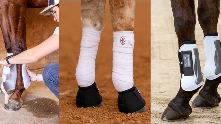
 Better diagnostics such as ultrasound or MRI can reveal lesions within the tendon sheath. Photo by Heather Smith Thomas
“In most windpuffs, there are no bony abnormalities. Whether you need to take radiographs would depend on how long the leg has been affected,” Baxter says. “Often radiographs can be completely normal yet the horse will still have fluid distension in the tendon sheath because it’s just a soft tissue injury in that part of the leg.”
The veterinarian would determine, on a case-by-case basis, whether radiographs might be helpful.
“This is an area we can also use MRI, if necessary,” Baxter says. “We can do a pretty good job with ultrasound and tenoscopy for diagnosis, but you could certainly document a lot more potential problems in the tendon sheath with an MRI. In a problematic case this could help us figure it out.”
For a valuable horse with an important career, MRI might be an option to consider to achieve the most accurate diagnosis.
“Some of these cases can be problematic, with chronic swelling, chronic tendonitis, and some of these horses can be quite lame. These cases would be the ones in which we’d use a more aggressive approach, such as tenoscopy,” Baxter says. “Whether or not you’d do an MRI before the surgery might depend on the case. The owner may not be able to afford to do both. If you have to do one or the other, I’d rather do the surgery, because then I can usually fix the problem at the same time I am looking inside the sheath with the arthroscope.”
If this approach didn’t resolve the issue, an MRI could be the next step, to find out more about what’s going on inside the leg. A CT scan may be used if access to MRI is unavailable. There are multiple options compared to what was available even 10 years ago, and treatment options are much better now than they were even five years ago.
“In the past we generally viewed windpuffs as not being very significant, but now we realize we have to look at them, to know if the fluid in the tendon sheath is telling us there is a problem there,” Baxter says. “In some cases we’d want to be more proactive in our diagnosing and preventing it from getting any worse.”
On rare occasions a horse might have problems with the smaller, digital annular ligament below the fetlock joint at the back of the pastern.
“This is further down. Instead of going across the back of the sesamoids at the back of the fetlock joint, this one is down in the pastern. This is reported to be a rare cause of lameness, but could be associated with windpuffs as well,” Baxter says as further proof it’s good measure to have the leg thoroughly checked.